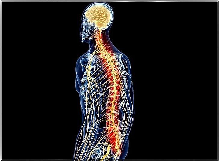
The Anatomy And Physiology Of The Spinal Cord
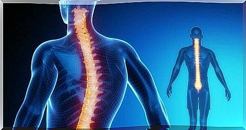
The spinal cord is part of the central nervous system, along with the brain. It runs from the back of the head to about the first vertebra.
There are 31 vertebrae connected to the spinal cord. It consists of a core of gray matter, where the neurons are located. This in turn is surrounded by white matter, where the axons are located.
Interestingly enough, on the one hand, the distribution of white and gray matter in the spinal cord is opposite to that in the brain. On the other hand, there are vertebrae, ligaments, ligaments, meninges, and cerebrospinal fluid that protect the spinal cord.
The spinal cord performs several functions. Among other things, it is responsible for receiving and processing (in a superficial way) information from the senses. However, these features are vital. An injury can have serious consequences, such as paralysis or loss of sensation.
The anatomy and physiology of the spinal cord
Grey dust
This gray matter is located in the inner part of the spinal cord. It is where the neurons are located and where the information is processed. It consists of several areas, namely: ventral, dorsal, lateral and intermediate.
- Dorsal horn: This part is responsible for information from the senses.
- Intermediate horn: The place where interneurons (or associative neurons), which connect neurons together, are found.
- Lateral Horn: Found only at the thoracic and lumbar levels. It is responsible for the homeostasis of the body and regulates the automatic nervous system.
- Ventral horn: This part is responsible for motor information.
In addition , this gray matter contains several nuclei with different functions:
- I-IV: responsible for the exteroceptive sensations. They store the sensations they receive from external stimuli, such as light.
- V-VI: responsible for the proprioceptive sensations. They pass on stimuli that are generated internally.
- VIII: carries out the transfer between mesencephalon and cerebellum. It is where the neurons from the mesencephalon take over to move to the cerebellum and vice versa.
- IX: the main motor area. It is where the neuronal bodies emerging from the motor cortex drive impulses of movement.
- X: Nucleus surrounding the central canal, containing glial cells, the supporting neurons.
The gray matter of the spinal cord is therefore primarily an exchange site for motor and sensory information. It must also quickly assess the information before it reaches its destination. The latter is important to activate the reflexes in emergency situations, such as when a painful stimulus enters.
White matter
The white matter of the spinal cord is where the axons, which send information up and down, are located. The most important function is again the transmission of information. Like the substantia nigra, it is also divided into several parts. In this case columns:
- Dorsal: transmits somatic information.
- Ventral and Lateral: The efferent pathways responsible for transmitting information from the brain to the muscles. They are also part of the motor system.
Within the white matter there are also different orbits – ascending and descending. These orbits are named after the two structures between which the information circulates. In addition, each job transmits different information.
These are the nerves:
- Gracilis and cuneatus: deal with the movements of the hands.
- Anterior and posterior spinocerebellar: unconscious movements of the muscles, joints and subcutaneous tissue.
- Spino olivar: It is unknown what exactly these orbits do.
- Spinothalamicus lateralis: painful and warm sensations.
- Spinotectal: information through spino-visual reflexes.
- Spinothalamic anterior: light touch and pressure.
- Anterior and lateral corticospinal : the smoothness and speed of movements.
- Tectospinal: movements by visual stimuli.
- Vestibulospinal: Responsible for maintaining balance.
- Olivospinal: affects the activity of motor neurons.
- Rubrospinal: deals with the activity of the extensor muscles.
The white matter is thus responsible for the transmission of motor and sensory information of a wide range of movements and sensations, communicating between different areas.
Ascending (sensory) nerve pathways
The ascending pathways, as the name implies, are responsible for transmitting the information gathered by the external senses (exteroceptive information) or internal stimuli (proprioceptive) to the brain. Further processing takes place there. Most of the ascending pathways terminate in the thalamus, except for the olfactory stimuli, which go directly to the forebrain.

They are ascending, centripetal, originate in the periphery and also provide information to higher centers. A number of nerve fibers serve as connections between different parts of the spine. In the same way others go to the upper centers connecting the spine and the brain.
They process information that may or may not reach consciousness.
In its simplest form , the pathway that reaches consciousness consists of three neurons. But there are also pathways that use more or fewer neurons. Many of the neurons in the ascending pathways go beyond that and are also part of the muscle reflexes.
It is those orbits that transmit information from somatic reflectors. There are two most common ways this information moves, namely:
- Nociceptive orbit: information about pain and temperature
- Sensory nerve pathway: information about superficial touch, proprioception and vibration.
Descending (motor) nerve pathways
These pyramidal tracts are the motor nerves that run down from the brain parts to the spinal cord. They are therefore responsible for conscious, fast, nimble, precise movements.
Three neurons are involved in the process of sending information to perform a movement. To do this, they follow this circuit:
- Neuron 1: Located in the prefrontal and premotor cortex.
- Neuron 2: does not always occur in this orbit. It is an interneuron or switching cell.
- Neuron 3: is located in the frontal horn of the spine.
All pyramidal tracts eventually show a contralaterality, which means that an injury in the right motor cortex, for example, causes an injury in the left part of the body.
The extrapyramidal system is responsible for unconscious movements, and arises from a subcortical structure, which pulls towards the spine. It thus regulates the performance of unconscious movements (such as walking, posture, muscle tone, alertness, and instinctive behavior).
Unlike the pyramidal system, it does not start in the cerebral cortex, but in the various subcortical structures.
Another important function of the descending nerves is to modulate the circuits that trigger reflexes in the spine. The adaptive capacity of the back reflexes can thus be modified according to behaviour.
Sometimes we need more force, at other times length or movement is needed, so that the movement adapts to the circumstances. Descending neural pathways are thus responsible for regulating these variables.
Spinal Reflexes
There are movements that we make unconsciously before the sensory information from the stimulus that causes the movement reaches our brain. These are reflexes, such as pulling your hand away from a hot surface, or closing your eyes to a loud noise. So we have no control over these movements.
The reflex is the simplest circuit in the nervous system. It starts in the receptors, structures that convert the energy of the stimuli into an electrical change in the afferent sensory nerves. These then send the stimuli to the integration center, the interneuron. Then the information is passed on to the efferent motor nerves, so that the muscle carries out the movement.
These movements are caused by the reflex arc. The soma of the neuron is located in the dorsal root ganglion and then passes through the dorsal root, where it communicates with the interneuron. That’s the neuron that integrates the information and passes it on to the motor neuron in the ventral horn, then exits the ventral root and sends the nerve impulse to the muscle for its contraction.
Bibliography
Carlson R. (2014). Fisiología de la conducta . Madrid, Espana: Pearson educación
Tortora GJ, Derrickson B.(2013). Principles of anatomy and physics . Buenos Aires: Editorial Medica Panamericana
Kandell ER, Schwartz JH y Jessell TM(2001). Principles of Neurociencia . Madrid: McGraw-Hill/Interamericana.